Elizabeth L. Brainerd and Sabine Moritz
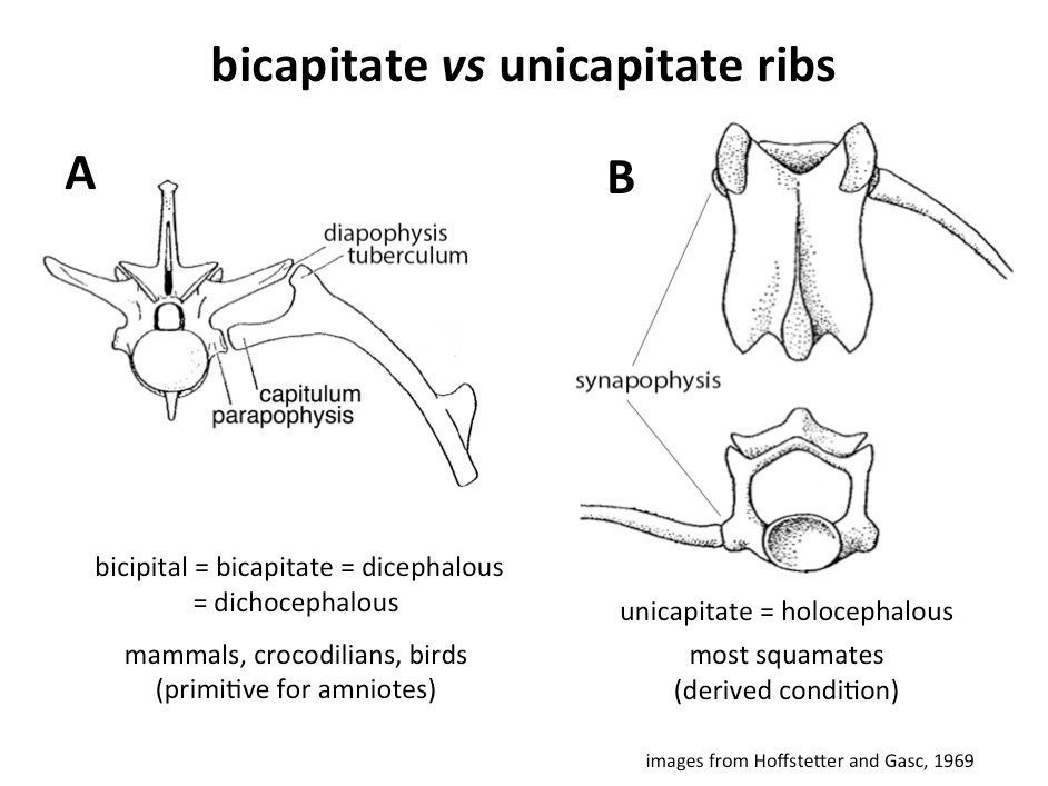
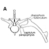 The joints between ribs and vertebrae vary widely among amniotes. The primitive condition for amniotes is bicapitate ribs that form two separate joints with the vertebrae (Figure 1A). The capitulum of the rib articulates with the parapophysis on the vertebral centrum, and the tuberculum articulates with the diapophysis on the transverse process. Squamates show a derived condition of unicapitate ribs that articulate with the vertebrae via single, hemispheric joints (Figure 1B). The single rib facet (synapophysis) on squamate vertebrae represents the fusion of the parapophysis and diapophysis, rather than the loss of one of them.
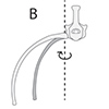 Motions of the ribs at the costo-vertebral joints contribute to lung ventilation in all amniotes except turtles. During breathing, the potential rotations of ribs at the costo-vertebral joints are described as rotation about three body axes: caliper rotation about a longitudinal axis (Figure 2A); bucket-handle rotation about a dorsoventral axis (Figure 2B); and pump-handle rotation about a mediolateral axis (Figure 2C).
Here we use X-ray Reconstruction of Moving Morphology (XROMM) to quantify the 6 DOF motions at costovertebral joints in non-serpentine squamates ("lizards"), a bird (Turkey) and a crocodilian (Alligator; Movie 1). In green iguanas, rib motions are strongly dominated by bucket-handle rotation during breathing, despite the potential for a wider range of rotations at the nearly hemispheric, unicapitate costovertebral joints.
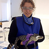
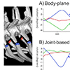 In crocodilians and birds, the double articulations between ribs and vertebrae should restrict motion to a single axis running through the parapophysis and diapophysis (Figure 1A). In Alligator mississippiensis (Figure 3), as expected, breathing is driven primarily by the hepatic piston. But the ribs do rotate and contribute to volume change (Movie 2). XROMM analysis shows that motion is indeed restricted primarily to one axis. When a joint coordinate system (JCS) is aligned to the body planes, rotations are around two axes, bucket and pump handle (Figure 4A). But when the axis is aligned to the costovertebral joint, running through the parapophysis and diapophysis, virtually all the motion can be captured as rotation about that joint (Figure 4B).

 The joints between ribs and vertebrae vary widely among amniotes. The primitive condition for amniotes is bicapitate ribs that form two separate joints with the vertebrae (
 Motions of the ribs at the costo-vertebral joints contribute to lung ventilation in all amniotes except turtles. During breathing, the potential rotations of ribs at the costo-vertebral joints are described as rotation about three body axes: caliper rotation about a longitudinal axis (
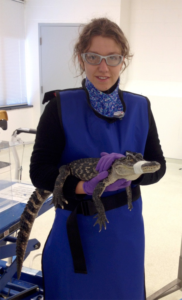
 In crocodilians and birds, the double articulations between ribs and vertebrae should restrict motion to a single axis running through the parapophysis and diapophysis (