Fish Bite Force

Gape-specific bite force and prey-size specific predator performance in the snail-eating black carp

N.J. Gidmark1*, N. Konow1, E. LoPresti2, and E.L. Brainerd1

  1. Brown University, Providence, RI, USA
  2. The University of California at Davis, Davis, CA, USA

*Author for correspondence: Nicholas J. Gidmark | Published article | Press release

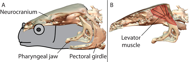
In this study we used X-ray Reconstruction of Moving Morphology (XROMM) to examine biting performance in a snail-eating carp species, the black carp (Mylopharyngodon piceus). Instead of using its oral jaws for crushing, black carp use the pharyngeal jaw apparatus, in the back of the throat (Figure 1). Using food items of known size and strength (Figure 2), we showed prey-size specific performance of the carp (Figure 3). The strongest items were only crushable at intermediate length, whereas weaker food items were crushable at a broader range of sizes. Next, we used XROMM to measure jaw and skull positions during in vivo biting on these same food items. From these XROMM animations, we measured muscle fiber lengths. Finally, we conducted in situ tests where we electrically stimulated the jaw-closing muscles and directly measured muscle force while using XROMM to measure bone posture (and, therefore, muscle length, Figure 4). By combining these three data streams (prey feeding trials, XROMM recordings of in vivo biting, and in situ recordings of maximum bite force, Figure 5), we demonstrated that: 1) Crushing performance in black carp is prey-size specific; 2) Prey size influences the instantaneous length of the jaw-closing muscle in black carp; and 3) bite force changes as a result of muscle length change, and this force contributes directly to bite performance. These results demonstrate how fundamental performance metrics of muscle fibers can function to limit performance on the organismal scale in black carp.

Pictures

Fig. 1: Oral jaws are not used for snail crushing in black carp; instead, the pharyngeal jaws function in fracturing snails. These jaws are modified fifth gill arches, which no longer have respiratory function; arches 1-4 are still normal gills. The pharyngeal jaw lies immediately anterior and medial to the pectoral girdle and is supported by a sling of muscles with no jaw joint. The main focus of this study was the levator muscle, which elevates the jaws into occlusion with the base of the skull. Fig. 2: We manufactured a series of different prey items by taking four different sizes of ceramic tubes (sizes, in millimeters, are indicated on the figure). Each size of prey item had a characteristic strength, as determined by a materials testing machine. We coated each prey item with polyurethane to increase its strength, and successive coats yielded at successively higher strengths. Using these manufactured Fig. 3: Each of our three individuals could fit all four sizes of prey between the jaws, and attempted to break them when they were filled with food. Each individual was given 5-10 attempts to crush each combination of sizes/strengths. Black-filled circles indicate 100% success in crushing the tubes of that size/strength, and success rates decline as the colors get lighter. White indicates zero success on a given food item. Note how performance varies not only with force (along the Y axis), but also with prey size (along the X axis). If an animal was unable to crush all of the food items of a given size and strength, we did not test stronger food items of that same size. Fig. 4: After the performance tests, we measured muscle force at a variety of muscle lengths. Using this setup, we electrically stimulated the muscle, recorded jaw and skull position using XROMM, and measured force using a force transducer. Since we had some feeding trials also within view of the X-ray machines, we were able to reconstruct what muscle length was used in vivo, and how that corresponded to muscle lengths along the force-length curve in situ. Fig. 5: By combining our in vivo and in situ data, we were able to show that bite performance in vivo is constrained by the force-length curve of the jaw adductor muscle. This graph is standardized to maximal muscle force for a given individual (Y axis) and the relative muscle length (X axis), where 1.0 is the muscle length at peak force. The grey symbols (circles, squares, and diamonds) represent in situ muscle lengths and forces from the apparatus in Fig. 4.The triangles represent the muscle length and strength of the weakest uncrushable food items of a given size (downward-pointing triangles) and the strongest crushable food items of a given size (upward-pointing triangles). Muscle lengths were measured in vivo while the animals crushed ceramic tubes, and forces were taken from Fig. 2.

Movie

Fish Bite Force: X-ray animation sequence: In live X-ray video, a black carp manages to crush a tube full of food deep in its mouth. The size of the tube has a major influence on the strength of its bite. The video also shows a computer animation of its jaw bones.

Reference

Gidmark, N.J., N. Konow, E. LoPresti, and E.L. Brainerd. (2013). Bite force is limited by the force-length relationship of skeletal muscle in black carp, Mylopharyngodon piceus. Biology Letters, 9:20121181. Published article. Press release.