
Fish Feeding
Biomechanics of jaw protrusion in common carp
- Brown University, Providence, RI, USA
- The George Washington University, Washington D.C., USA
In this study we are using X-ray Reconstrucion of Moving Morphology (XROMM) to examine the movements of several oral jaw bones and understand the mechanics of jaw protrusion in common carp, Cyprinus carpio.
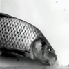
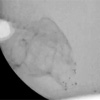
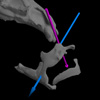
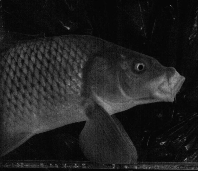
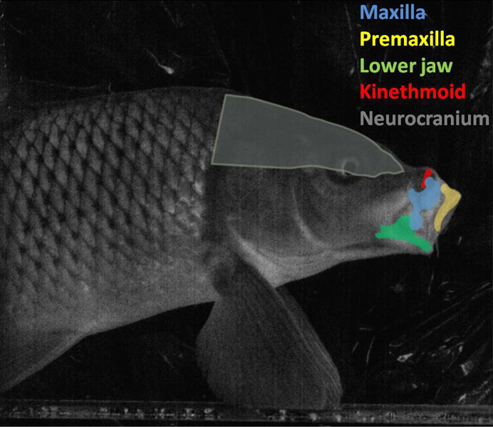
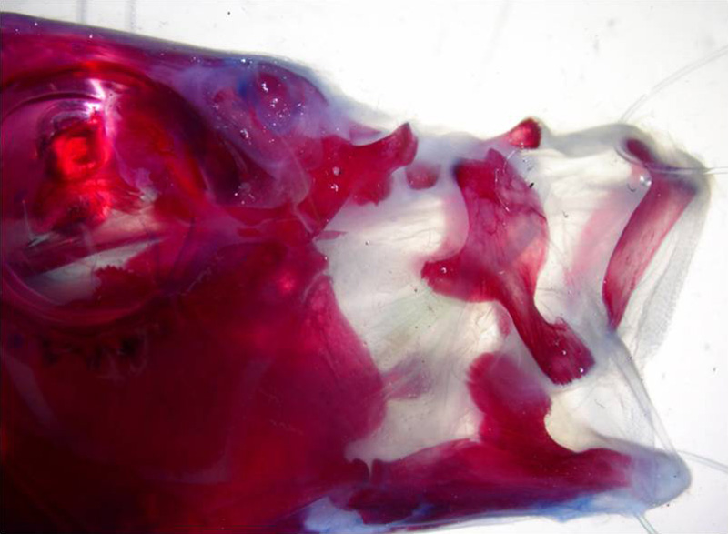
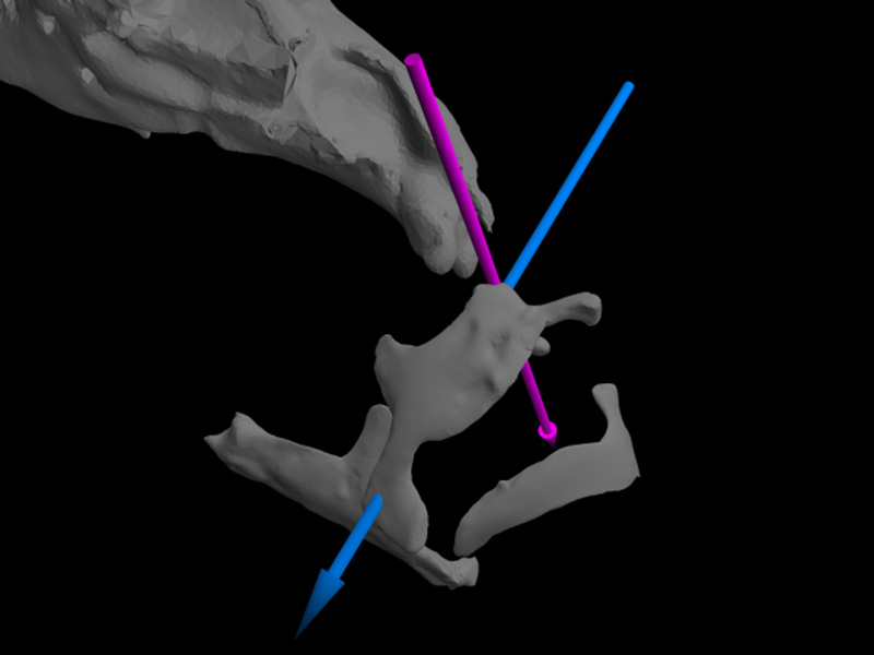
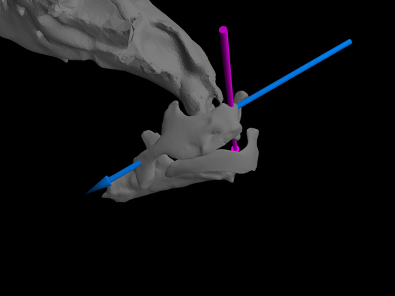
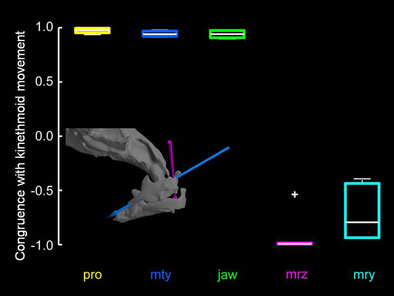
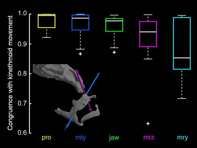
Cypriniform fishes (commonly known as minnows, carps, goldfish, koi and loaches) are capable of impressive protrusion of the oral jaws (Figure 1). This protrusion can be observed in conjunction with or separate from opening of the mouth, likely resulting in nuanced control of water flows within the oral and pharyngeal cavities (Movie 1). This control is pivotal for the bottom-feeding lifestyle of common carp. One morphological oddity that can be seen in common carp (and in all cypriniform fishes except three species, which have secondarily lost it) is the presence of the kinethmoid, a highly mobile midline bone (Figure 2) located in a ligamentous meshwork supported by upper jaw bones and the neurocranium. Because of its location in ligaments and occluded by other bones, even anatomical preparations aimed at demonstrating bone may not show it clearly (Figure 3). Because of these limitations, previous in-vivo studies have been unable to explicitly examine its movements or the mechanics of how the upper jaws are protruded in kinethmoid-bearing species. We used marker-based (Figure 4) XROMM to create precise and accurate animations of 3D oral jaw bones and their motions during jaw protrusion in suction-feeding carp (Movie 3) based on x-ray videos (Movie 2). We used anatomical axes to track specific anatomically relevant degrees of freedom in these bones during both open-mouth (Figure 5) and closed-mouth (Figure 6) protrusion events. Kinematic curves for the ventral translation of the maxilla, premaxillary protrusion, and lower jaw rotation are all three highly correlated (based on a cross correlation analysis without lag) with kinethmoid rotation in both open-mouthed (Figure 7) and closed-mouth (Figure 8) protrusion events. Since the lower jaw is the only one of those movements that is less pronounced in closed-mouth trials, we hypothesize that muscles attaching to the maxilla (specifically A1beta) drive the maxilla to translate ventrally. We further hypothesize that maxillary translation causes kinethmoid rotation and ultimately premaxillary protrusion. These findings are significant for two reasons. First, we have experimental evidence indicating how this novel mechanism of jaw protrusion functions biomechanically. Second, and possibly most important, we have empirically shown decoupling of upper jaw protrusion from lower jaw abduction. Most fishes that protrude the upper jaw do so by abducting the lower jaw. The decoupling seen in common carp is likely a derivation that allows for precise control of water flows within the mouth and thus great dexterity in sorting food particles from the substrate where these fishes feed.
Pictures

Movies