Soft Tissue Artifact

Comparing knee kinematics between XROMM and traditional optical motion capture during a jump-cut maneuver

D.L. Miranda1,2, M.J. Rainbow1,2, J.J. Crisco1,2,3, and B.C. Fleming1,2,3

*Author for correspondence: Braden C. Fleming | Published article

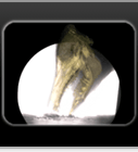
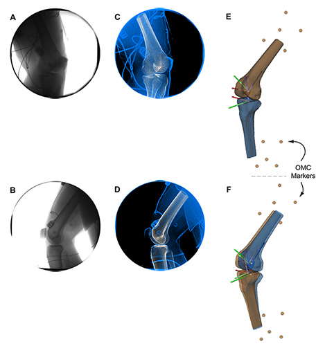
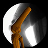
In this study we compared the kinematic measurements obtained using X-ray Reconstruction of Moving Morphology (XROMM) and a traditional optical motion capture system while male and female subjects performed a jump-cut maneuver. Many laboratories investigate jumping and cutting activities in order to better understand the biomechanics associated with non-contact ACL injury. Traditional optical motion capture is widely used; however, it is subject to soft tissue artifact (STA). XROMM offers a unique approach to collecting skeletal motion without STA (Movie 1). Therefore, the goal of this study was to compare how STA affects the six-degrees-of-freedom motion of the femur and tibia during a jump-cut maneuver associated with non-contact ACL injury. We recruited ten volunteers and asked them to perform a jump-cut maneuver while their landing leg was imaged using XROMM and a traditional optical motion capture system. We compared the within-bone motion differences using anatomical coordinate systems for the femur and tibia, respectively (Figure 1). We also compared the knee joint kinematic measurements during two periods: before and after ground contact. We observed that, over the entire activity, the within-bone motion differences between the two motion capture techniques were significantly lower for the tibia than the femur for two of the rotational axes (flexion/extension, internal/external) and the origin. We also observed that the knee joint kinematics obtained from both systems were in best agreement before landing (Figure 2). Not surprisingly, we observed the kinematic deviations between the two techniques increase significantly after contact. In conclusion, this study provides information on the kinematic discrepancies between XROMM and traditional optical motion capture that can be used to optimize methods employing both technologies for studying dynamic in vivo knee kinematics and kinetics during a jump-cut maneuver.

Pictures

Panels A and B represent a single frame of the biplanar videoradiography data for source 1 and 2, respectively. Panels C and D represent the same frame of the biplanar videoradiography data (blue) after image processing. The 3-D models of the tibia and femur driven by optical motion capture (tan) and biplanar motion capture (blue) are shown in panels E and F. All four independently tracked anatomical coordinate systems are also shown. The short and lighter coordinate systems are being driven by OMC and the long and darker coordinate systems are being driven by biplanar videoradiography. The external markers for the thigh and shank are also shown in tan. Panel E represents the initial frame, where OMC and biplanar videoradiography are perfectly aligned. Panel F represents a frame where soft tissue artifact is affecting the OMC driven bones and coordinate systems. Example OMC (dotted red) and biplanar videoradiography (solid green) knee flexion angle and ground reaction force (solid blue) data versus time. The dotted vertical lines represent the contact event. The period before contact is period A and the period after contact is period B. Period A began and period B ended when the femur and tibia entered and exited the FOV of the biplanar videoradiography system. Thus, the arrows shown above the ME/LA translation graph denote the time point where the knee entered and exited the field of view of the biplanar videoradiography system, respectively.

Movie

Jump Cut: X-ray video & XROMM animation

Reference

Miranda, D.L., M.J. Rainbow, J.J. Crisco, and B.C. Fleming (2013). Kinematic differences between optical motion capture and biplanar videoradiography during a jump-cut maneuver. Journal of Biomechanics. 46(3): 567-573. Published article.

Related Publications

Miranda, D.L., P.D. Fadale, M.J. Hulstyn, R.M. Shalvoy, J.T. Machan, and B.C. Fleming. (2012). Knee Biomechanics during a Jump-Cut Maneuver: Effects of Gender and ACL Surgery. Medicine and Science in Sports and Exercise. [Epub ahead of print].
Published article.

Miranda, D.L., J.B. Schwartz, A.C. Loomis, E.L. Brainerd, B.C. Fleming, and J.J. Crisco. (2011). Static and dynamic error of a biplanar videoradiography system using marker-based and markerless tracking techniques. Journal of Biomechanical Engineering. 133(12): 121002. Published article.

Miranda, D.L., M.J. Rainbow, E.L. Leventhal, J.J. Crisco, and B.C. Fleming. (2010). Automatic determination of anatomical coordinate systems for three-dimensional bone models of the isolated human knee. Journal of Biomechanics. 43(8): 1623-1626.
Published article.

Author Affiliations

1Department of Orthopaedics, The Warren Alpert Medical School, Brown University
and Rhode Island Hospital, Providence, RI, USA

2Center for Biomedical Engineering, Brown University, Providence, RI, USA

3School of Engineering, Brown University, Providence, RI, USA