

Hardware
Two types of high-speed, biplanar x-ray video systems are in general use today: systems based on mobile C-arm fluoroscopes and custom-built biplanar x-ray rooms.
The advantage of mobile C-arm fluoroscopes is the relatively low cost of refurbished units (less than $200,000 for a biplanar system, including high-speed video cameras). The main disadvantages are low tube current (fluoroscopic levels, generally 20 mA maximum) and the physical constraints imposed by the C-structures.
The main advantages of a custom-built, biplanar x-ray room are better image quality due to higher tube currents (radiographic rather than fluoroscopic levels), a larger imaged volume, and more open space within the apparatus for experimental equipment and research subjects.
Mobile C-arm Fluoroscopes
 In the past, the high cost of cineradiographic equipment has limited the number of single plane systems dedicated to zoological work to a small handful, and no biplanar systems were available. The relatively low cost of refurbished C-arm fluoroscopes should now make it possible for more research groups in comparative biomechanics to acquire biplanar videofluoroscopy hardware. It is our hope that C-arms, combined with the methods that we have developed for marker-based XROMM and manual markerless XROMM (i.e. Scientific Rotoscoping), will make it possible for the potential power of XROMM analysis in field of comparative musculoskeletal biomechanics to be realized.
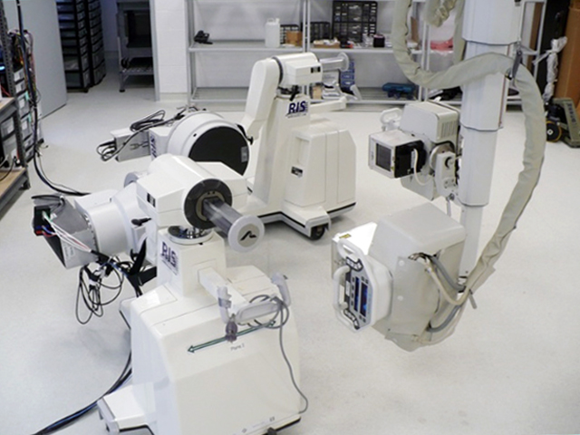
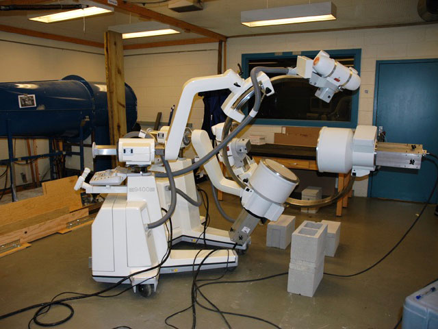 We have found that two mobile C-arm fluoroscopes, retrofitted with 30-cm image intensifiers and high-speed video cameras, form an effective system for measuring accurate 3D skeletal kinematics of small to medium-sized animals. Some suitable, relatively inexpensive models are the OEC 9000 and OEC 9400, which were originally manufactured by OEC Medical Systems in the early 1990s. These OEC models come standard with 23-cm (9") image intensifiers (IIs), which is inconveniently small for biplanar capture of movement. These old C-arms can be retrofitted with new 30-cm (12") IIs and high-speed video cameras.
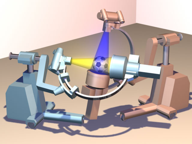
Radiological Imaging Services, Hamburg PA (800-748-2040) has experience refurbishing and retrofitting C-arms for high-speed imaging. In this process, the x-ray source is centered over a new 30-cm II and the standard camera and video monitors are removed and replaced with a user-specified high-speed video system. However, even with 30-cm IIs, the imaged volume is quite small — about the size of a standard soccer ball.
Biplanar x-ray rooms
 In a custom-built biplanar videoradiography room, the x-ray equipment and physical layout can be designed specifically for the intended research projects. Compared with C-arms, there is the potential for larger IIs, higher tube currents, and more flexible positioning of the x-ray tubes and IIs. The disadvantage, relative to C-arms, is cost. The cost for the high-speed video cameras, x-ray generators, x-ray tubes, IIs, and gantries to position the equipment is expected to be in the range of $800,000 to $1M (for a biplanar system with high-speed pulsed x-ray generation, 16" (40cm) image intensifiers, and high-end video cameras). The need to renovate a large room with necessary ceiling supports for the gantries and x-ray shielding also adds substantial cost. We recommend a room that is at least 20'x20' (6m x 6m); the W.M. Keck Foundation XROMM Facility at Brown University is 21'x33'.
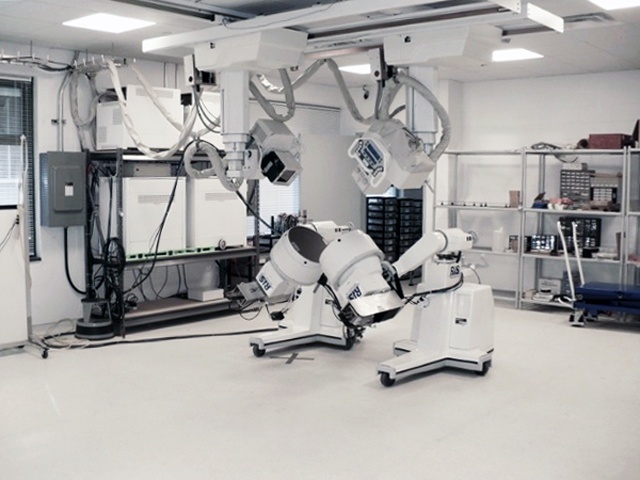
 The W.M. Keck Foundation XROMM Facility biplanar room at Brown University consists of two Varian model G-1086 x-ray tubes, two EMD Technologies model EPS 45-80 pulsed x-ray generators, two Dunlee model TH9447QXH590 image intensifiers (16" diameter), and two Phantom v10 high-speed digital video cameras. The x-ray tubes are suspended from the ceiling by tube cranes and the IIs are mounted on mobile gantries. The components are set up such that the two x-ray beams intersect each other close to the IIs. A treadmill, trackway or other animal support/enclosure is placed such that the research animal performs the behavior of interest (running, jumping, flying) in the volume where the x-ray beams intersect.
The Brown University system can deliver pulsed x-ray generation at up to 100 Hz, and can record in continuous x-ray generation mode at up to 1000 fps. With the Phantom v10 cameras, the pixel resolution is 1800x1800. Overall resolution of the imaging chain is about 2 line pairs/mm. Radio-opaque beads can be tracked to within ±0.1 mm in 3D space. The x-ray system was designed and integrated by Marty Kulis of Imaging Systems and Service, Painesville OH (mkulis@issi-na.com; 440-724-8002). The mobile gantries supporting the IIs were designed and built by Ryan Reeser at Radiological Imaging Services, Hamburg PA (800-748-2040).