
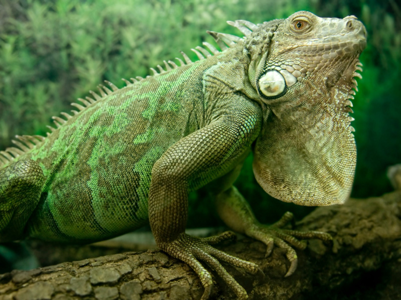
Iguana Breathing
Rib kinematics & intercostal muscle strain during breathing in Iguana iguana
Published article (PDF,74KB).
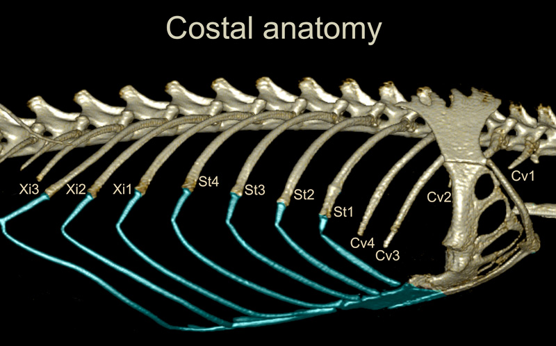
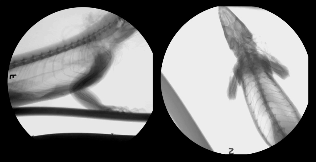
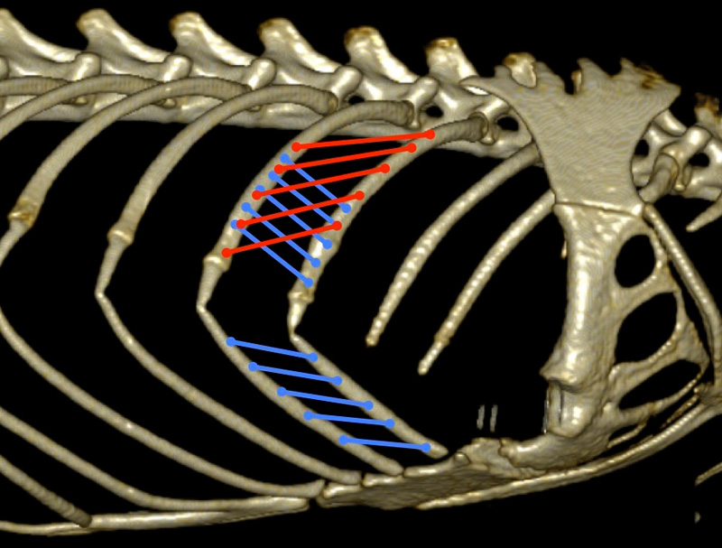
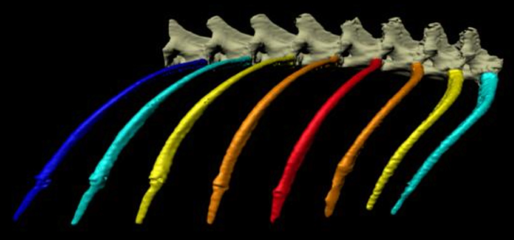
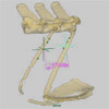
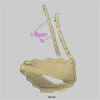
Marker-based and markerless X-ray Reconstruction of Moving Morphology (XROMM) were used to create precise and accurate animations of 3D rib motion in green iguanas during deep breathing. Muscle fascicle strain was measured by mapping the attachment points of external intercostal (EI) and internal intercostal (II) muscle fascicles onto the 3D rib models (Figure), and running the XROMM animations to measure the change in distance between the attachment points (Movie). Green iguanas have four cervical (Cv) ribs, four sternal (St) ribs that articulate with the sternum via long costal cartilages, and three xiphisternal ribs (Figure). XROMM analysis shows that the dorsal osseous ribs and ventral costal cartilages move as separate rigid elements, connected by thin cartilaginous regions that act as joints (Movie). XROMM animations reveal little movement of Cv1 and Cv2, increasing amounts of rotation in Cv3-St1, the greatest rotation in St2, and gradually decreasing rotation in St3-4 and more caudal ribs. In the intercostal space between the osseous portions of St1 and St2, the EI lengthen during exhalation and shorten during inhalation, whereas between St3 and St4, the EI shorten during exhalation and lengthen during inhalation. The costal cartilages show no craniocaudal rotation gradient, but they fold back during exhalation such that the parasternal II fibers lengthen during exhalation and shorten during inhalation (Movie). These results are consistent with the previously described EMG patterns of the EI and II in iguanas (Carrier, 1989), and are strikingly similar to recent consensus developing on the functions of EI and parasternal II in dogs (Carrier, 1996; De Troyer et al., 2005).
Pictures
Movies
References
Carrier D.R. (1989). Ventilatory action of the hypaxial muscles of the lizard Iguana iguana: a function of slow muscle. Journal of Experimental Biology. 143: 435–457.
Carrier D.R. (1996). Function of the intercostal muscles in trotting dogs: ventilation or locomotion? Journal of Experimental Biology. 199: 1455–1465.
De Troyer A., Kirkwood P.A., and Wilson T.A. (2005). Respiratory action of the intercostal muscles. Physiological Reviews. 85: 717–756.