About XROMM
X-ray Reconstruction of Moving Morphology (XROMM) is a 3D imaging technology, developed at Brown University, for visualizing rapid skeletal movement in vivo.
XROMM combines 3D models of bone morphology with movement data from biplanar x-ray video to create highly accurate (±0.1 mm) re-animations of the 3D bones moving in 3D space.
Rapid bone motion, such as during bird flight, frog jumping, and human running, can be visualized and quantified with XROMM.
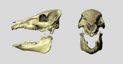
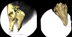
3D models of pig skull and lower jaw from a CT scan. XROMM requires independent models of each bone for re-animation. Models created in Amira, cleaned in Geomagic, and animated in Autodesk Maya. Animation by E. Brainerd.
X-ray video setup
Biplanar x-ray video data collection for a study of mastication in minipigs. Two C-arm fluoroscopes retrofitted with high-speed video cameras collect x-ray video from two perspectives. The resulting images can be seen on the computer monitors in the background. Video by E. Brainerd.
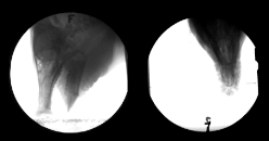
Minipig mastication in lateral x-ray projection. Bones appear dark and air appears white in this x-ray positive movie. Video was recorded at 250 frames per second and is played back here at approximately 1/4 real speed. Video by K. Metzger.
Minipig mastication in ventro-dorsal x-ray projection. Bones appear dark and air appears white in this x-ray positive movie. Video was recorded at 250 frames per second and is played back here at approximately 1/4 real speed. Video by K. Metzger.
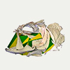
Precise re-animation of the CT bone models to match the movement recorded in the x-ray movies. In this study, small metal markers were used to determine the correct pose and position of the bones in 3D space (to within ±0.1 mm). Maya animation by D. Baier.
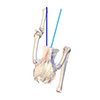
Precise re-animation of the CT bone models to match the movement recorded in the x-ray movies (ventro-dorsal view). Note that the whole bone does not have to be in view to reconstruct the position of every point on a rigid bone. Maya animation by D. Baier.
Upcoming Short Course
2016 Summer Short Course in X-ray Reconstruction of Moving Morphology (XROMM)
June 6-10, 2016
Brown University
Department of Ecology & Evolutionary Biology
Providence, RI, 02912, USA
This one-week course is designed for faculty, postdocs, and graduate students who are interested in using XROMM in the field of comparative biomechanics. The course will provide hands-on instruction in marker-based and markerless XROMM animation, analysis of 3D skeletal kinematics, data management and measurement of precision and accuracy. The course is funded by an NSF Advances in Biological Informatics grant.
To apply complete the Google application form. Review of applications will begin on March 21, 2016 and continue until the course is filled.
Project Showcase
- Fish Suction Power
- Swimming muscles power suction feeding in largemouth bass
In this study we used X-ray Reconstruction of Moving Morphology (XROMM) to measure the power required for suction feeding in largemouth bass (Micropterus salmoides), and compare it to the power available from cranial and body muscles.
- Long-Axis Rotation
- A missing degree of freedom in avian bipedal locomotion
In this study we are using X-ray Reconstruction of Moving Morphology (XROMM) to measure the 3-D motion and forces/moments in a chicken-like bird, the Helmeted Guineafowl (Numida meleagris), during maneuvering and steady locomotion.
- Jump Cut
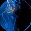
- Biomechanics of male and female ACL-intact and ACL-reconstructed athletes during a jump-cut maneuver
In this study we used X-ray Reconstruction of Moving Morphology (XROMM) to compare kinematic and kinetic knee measurements during a jump-cut maneuver.
In the News
NSF Science Nation Special Report
XROMM puts biomechanics on the fast track
New biomechanics visualization technology can be shared
among scientists in open source database
Bass use body's swimming muscles to suck in food
Posted Jun. 22, 2015 on news.brown.edu. Story by David Orenstein.
XROMM Project: Fish Suction Power
The birth of a dinosaur footprint: Subsurface 3D motion reconstruction and discrete element simulation reveal track ontogeny
Published Dec. 8, 2014 in PNAS early edition
Covered by: livescience.com | phys.org/news | sciguru.org | pfalkingham.wordpress.com
Visualizing rapid skeletal movements
Posted Sep. 20, 2013 on research.gov
XROMM Projects featured: Avian Bipedal Locomotion | Pig Feeding
Also mentioned on twitter by @NSF
Size of lunch dictates force of crunch
Posted Feb. 12, 2013 on news.brown.edu. Story by David Orenstein.
XROMM Project: Fish Bite Force
Additional coverage by: news.science360.gov | eurekalert.org | sciencedaily.com | ria.ru/science | scienceblog.com | redorbit.com | nsf.gov/news | esciencenews.com | phys.org/news
Funding Acknowledgements
We thank the Office of the Vice President for Research at Brown University, the RIH Orthopaedic Foundation, and the Bushnell Research and Graduate Education Fund for essential seed funding at the start of the XROMM development project. The W.M. Keck Foundation generously provided funding for the development of biplanar videoradiography hardware, and in support of our interdisciplinary collaborative development of XROMM software. The Instrument Development for Biological Sciences Program at the US National Science Foundation provided funding for the development of low-cost x-ray hardware and XROMM software for comparative biomechanics research.
Some material on this web site is based on work supported by the National Science Foundation. Any opinions, findings, and conclusions or recommendations expressed in this material are those of the author(s) and do not necessarily reflect the views of the National Science Foundation.